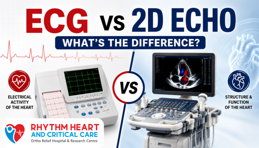
Heart tests play a major role in detecting cardiac problems early and preventing serious complications. Many patients often get confused between ECG vs 2D Echo: What’s the Difference?
This is because both tests are commonly used to diagnose the heart. While an ECG records the heart’s electrical activity, a 2D echo uses ultrasound to create a live image of the heart’s structure and blood flow.
At Rhythm Heart & Critical Care Hospital, experienced cardiologists use advanced diagnostic technologies to evaluate heart health accurately and provide the Appropriate Treatment for Patients treatment for patients suffering from chest pain, breathlessness, irregular heartbeat, or high blood pressure.
ECG vs 2D Echo: Knowing the Key Difference
Comparison of ECG and 2D echo: What is the difference?It is important to know that the two tests have different purposes in cardiac diagnosis. An ECG (electrocardiogram) checks the heart’s electrical rhythm and can help identify irregular heartbeats, heart attacks, or conduction problems.
On the other hand, a 2D echo evaluates the heart’s structure, valves, chambers, and pumping function through ultrasound imaging. Both tests are painless, non-invasive, and commonly performed in cardiac care hospitals for early detection of heart disease and better treatment planning.
ECG vs 2D Echo: Major Differences Explained
| Factor | ECG (Electrocardiogram) | 2D Echo (Echocardiography) |
| Purpose of the Test | Evaluates electrical activity and heart rhythm | Examines heart structure, valves, chambers, and blood flow |
| Technology Used | Uses skin electrodes to record electrical impulses | Uses ultrasound waves to create live heart images |
| Detection of Heart Problems | Detects arrhythmias, heart attacks, and rhythm disorders | Detects valve disease, heart enlargement, weak heart muscles, and congenital defects |
| Imaging Capability | Does not provide heart images | Provides detailed real-time heart images |
| Procedure Duration | Usually completed within 5–10 minutes | Takes around 20–40 minutes |
| Pain and Safety | Painless, non-invasive, and safe | Painless, radiation-free, and non-invasive |
| Role in Emergency Diagnosis | Quickly identifies heart attacks and rhythm issues | Evaluates heart function and damage after stabilization |
| Evaluation of Heart Valves | Cannot directly assess heart valves | Effectively detects valve leakage or narrowing |
| Monitoring Heart Function | Tracks electrical rhythm changes over time | Monitors pumping function and blood flow |
| Importance in Cardiac Care | Important for quick rhythm assessment | Essential for detailed structural heart evaluation |
1. Purpose of the Test
ECG and 2D echo are important heart tests that evaluate different aspects of heart health. ECGs primarily focus on electrical activity and heart rhythm, and can help doctors detect irregular heartbeats, arrhythmias, or signs of a heart attack.
A 2D echo, however, provides detailed images of the heart’s structure, valves, chambers, and blood flow. Together, these tests help cardiologists diagnose and manage various cardiac conditions accurately and effectively.
2. Technology Used
ECG uses small electrodes attached to the chest, arms, and legs to record the electrical impulses generated by the heart. These impulses are displayed as wave patterns that help doctors identify rhythm abnormalities.
In contrast, 2D echo uses high frequency ultrasound waves to create vivid, moving images of the heart. This technology allows cardiologists to observe heart muscles, valve movement and blood flow in real time, making it very useful for advanced cardiac diagnostics and treatment planning.
3. Detection of Heart Problems
An ECG is commonly used to identify irregular heartbeat, heart rhythm disturbances, and signs of previous or ongoing heart attacks. It provides quick results and is especially useful in emergencies.
2D echo detects structural heart problems such as enlarged heart chambers, valve abnormalities, congenital heart defects, blood clots, and weakened heart muscle. By combining both tests, doctors can better understand a patient’s heart condition and recommend the most appropriate treatment.
4. Imaging Capability
One of the major differences between ECG and 2D echo is the image processing power. The ECG does not produce an actual image of the heart. Instead, electrical activity is recorded as a graphic waveform on a monitor or paper.
2D Echo uses ultrasound technology to create detailed images of your heart in real time. These videos allow physicians to assess myocardial contraction, valve function, ventricular size, and blood flow patterns, making them important diagnostic tools in modern cardiology and cardiovascular disease management.
5. Procedure Duration
An ECG is a quick diagnostic test that usually takes about 5 to 10 minutes. It is usually done during a routine checkup or an emergency test for chest pain or palpitations.
A 2D echo generally takes 20 – 40 minutes depending on the patient’s condition and the complexity of the examination. Since 2D echo involves detailed imaging of heart structures and blood flow, it requires more time for accurate cardiac assessment and diagnosis of underlying heart diseases.
6. Pain and Safety
Both ECG and 2D echo are completely safe, painless, and non invasive procedures widely used in cardiac diagnosis. Neither test involves surgery, needles, or radiation exposure, making them suitable for patients of all age groups, including elderly individuals and children.
ECG simply records electrical signals through skin electrodes, while 2D echo uses harmless sound waves to create heart images. Their safety and convenience make them essential tools for routine heart evaluation and long-term monitoring of cardiac health.
7. Role in Emergency Diagnosis
ECG plays a critical role in emergency situations because it delivers rapid information about heart rhythm abnormalities and possible heart attacks within minutes. Doctors often perform ECG immediately in patients with chest pain, breathlessness, or dizziness.
A 2D echo provides more detailed information about heart muscle damage, pumping strength, valve function, and blood flow after the patient is stabilized. Together, these tests help cardiologists make quick and accurate decisions during emergency cardiac care and treatment planning.
8. Evaluation of Heart Valves
ECG cannot directly visualize or evaluate heart valves, though it may sometimes show indirect signs of valve-related stress on the heart. A 2D echo is highly effective in examining valve structure and function in detail.
It helps detect conditions such as valve leakage, stenosis, infection, and abnormal valve movement. The Doppler technique used during 2D echo also assesses blood flow through the valve, making it one of the good tools for diagnosing and planning treatment for heart valve disease.
9. Monitoring Heart Function
Doctors usually use an electrocardiogram to monitor changes in the heart’s rhythm over time, especially in patients who have arrhythmia, a pacemaker, or have had a heart attack.
This helps monitor electrical activity and detect abnormal patterns early. 2D echo monitors the heart’s pumping capacity, ventricular size, muscle movement, and blood flow efficiency.
This test is extremely useful in evaluating heart failure, cardiomyopathy, and recovery after cardiac procedures. Regular monitoring with these tests helps ensure timely treatment and better long-term heart health management.
10. Importance in Cardiac Care
ECG and 2D echo are important diagnostic tools in modern cardiology and play an important role in the early detection of heart disease. The ECG provides valuable information about the heart’s electrical activity, and the 2D echo provides detailed visualization of the heart’s structure and function.
Cardiologists often use both tests together to make an accurate diagnosis and treatment plan. These studies can help identify serious heart disease early, improve patient outcomes, and support effective treatment of heart disease through prompt medical intervention and monitoring.
Which Test is Better – ECG or 2D Echo?
ECG and 2D echo are used for different purposes, so there is no single “good” test. While the ECG is great for checking the heart’s rhythm and electrical activity, the 2D echo provides detailed structural and functional information about the heart.
In many cases, doctors recommend both tests simultaneously to fully evaluate your heart disease and make an accurate diagnosis.
Conclusion
Knowing the difference between ECG and 2D echo helps patients make informed decisions about heart diagnosis and treatment. Early testing can identify hidden cardiac conditions before they become serious.
If you experience chest pain, breathlessness, dizziness, palpitations, or high blood pressure, consult the heart specialists at Rhythm Heart & Critical Care Hospital for advanced cardiac testing and expert heart care.
Book your ECG or 2D Echo appointment today and take the first step toward better heart health.
FAQ’s
1. What is an ECG test?
ECG is a test that records the electrical activity and rhythm of the heart.
2. What is a 2D echo test?
A 2D echo is an ultrasound-based imaging test that shows live images of the heart structure and blood flow.
3. Is ECG painful?
No, ECG is completely painless and non-invasive.
4. How long does a 2D echo take?
A 2D echo usually takes around 20 – 40 minutes depending on the examination.
5. Which test is better for heart blockage?
Both tests help in diagnosis, but additional tests like angiography may be needed to confirm blockages.
6. Can ECG detect valve problems?
ECG may show indirect signs, but a 2D echo is more accurate for evaluating heart valve conditions.
7. Is fasting required before ECG or 2D echo?
Usually, fasting is not required for standard ECG or 2D echo tests.
8. Why do doctors recommend both ECG and 2D echo?
Both tests provide different information and together offer complete heart evaluation.
9. Are ECG and 2D echo safe for elderly patients?
Yes, both tests are safe and commonly performed in elderly patients.
10. Where can I get ECG and 2D Echo in Nagpur?
You can visit Rhythm Heart & Critical Care Hospital for advanced cardiac testing and expert diagnosis.