- 19, Opp. Ramkrishna Math, Rahate Colony Road, Dhantoli, Nagpur -12, +91 9413564868
Peripheral Angiography
Peripheral angiography is a medical imaging test used to evaluate blood flow in the arteries outside the heart, particularly in the legs, arms, kidneys, and neck. This procedure helps detect blockages, narrowing, or abnormalities in the peripheral arteries, which can lead to conditions like Peripheral Artery Disease (PAD). By injecting a contrast dye into the blood vessels and capturing X-ray images, doctors can identify circulation problems and determine the best course of treatment.
Peripheral Angiography Procedure
Peripheral angiography is performed for various diagnostic and therapeutic purposes, including:
- Detecting Peripheral Artery Disease (PAD): Identifies narrowed or blocked arteries in the limbs, which can cause pain, numbness, and poor circulation.
- Evaluating Blood Flow Before Surgery: Helps assess arteries before procedures like bypass surgery or stent placement.
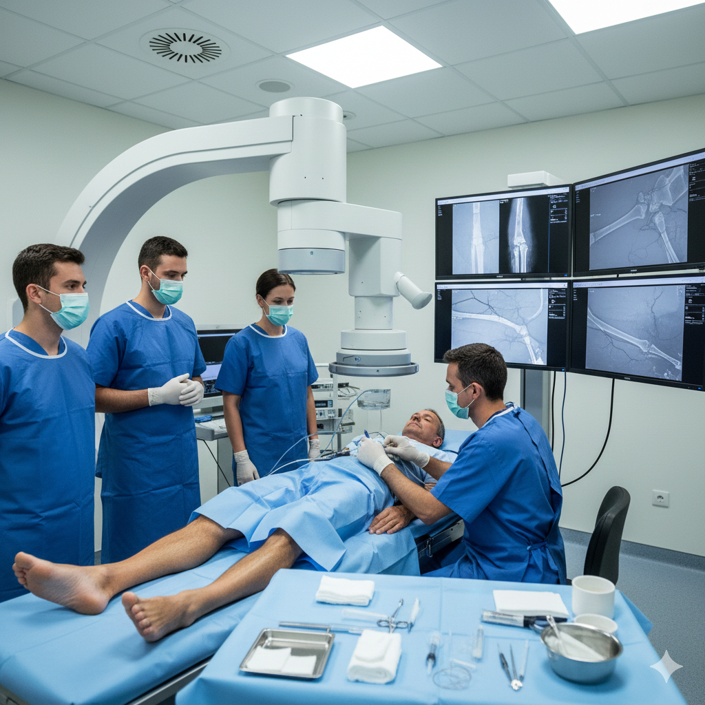
How is Peripheral Angiography Performed?
The procedure is typically conducted in a hospital or specialized vascular imaging center. The steps include:
1. Preparation:
- The patient may be asked to avoid food and drink for a few hours before the test.
- Blood tests and kidney function tests are conducted to ensure the body can handle the contrast dye.
- The groin or wrist area is shaved and sterilized to prepare for catheter insertion.
2. Procedure:
- A local anesthetic is administered to numb the area where the catheter will be inserted.
- A thin, flexible tube (catheter) is inserted into an artery, usually in the groin or wrist.
- Contrast dye is injected through the catheter, making the arteries visible on X-ray images.
- The doctor captures a series of X-ray images to observe blood flow and detect blockages.
3. Post-Procedure Care:
- The catheter is removed, and pressure is applied to the insertion site to prevent bleeding.
- The patient is monitored for a few hours to check for any complications.
- Drinking plenty of water helps flush the contrast dye from the body.
Risks and Complications
Although peripheral angiography is generally safe, some risks include:
- Allergic reaction to contrast dye
- Bleeding or bruising at the catheter insertion site
- Blood clot formation
- Kidney damage in patients with pre-existing kidney disease
- Infection (rare but possible)
What Happens After Peripheral Angiography?
Monitoring & Recovery
The patient is observed for a few hours to check for bleeding, allergic reactions, or complications from the contrast dye.
Hydration
Drinking plenty of fluids helps flush out the contrast dye from the body and supports kidney function.
Activity Restrictions
Patients are advised to rest and avoid strenuous activities for at least 24-48 hours to allow the catheter insertion site to heal.
Treatment Decisions
If blockages are detected, the doctor may recommend angioplasty, stent placement, or bypass surgery based on severity.
Medication & Lifestyle Changes
Blood thinners, cholesterol-lowering drugs, and lifestyle changes (healthy diet, exercise, and quitting smoking) may be prescribed to improve circulation.
Follow-Up Appointments
Regular check-ups are scheduled to monitor recovery and ensure proper blood flow in the treated arteries.