- 19, Opp. Ramkrishna Math, Rahate Colony Road, Dhantoli, Nagpur -12, +91 9413564868
2D Echo Medical Test
Advanced Heart Imaging for Accurate Cardiac Diagnosis
At Rhythm Heart & Critical Care Hospital, we provide advanced 2D/3D Echocardiography with Doppler services for detailed evaluation of heart structure, blood flow, and overall cardiac function. This non-invasive imaging test uses ultrasound waves to create high-quality real-time images of the heart, helping cardiologists diagnose various heart conditions with precision and accuracy.
Unlike standard imaging techniques, 2D/3D Echocardiography with Doppler offers a more comprehensive assessment by combining two-dimensional and three-dimensional heart imaging along with Doppler technology to analyze blood circulation through heart chambers and valves. This advanced diagnostic method plays a vital role in detecting heart valve disorders, congenital heart diseases, cardiomyopathy, heart failure, blocked arteries, and abnormal blood flow patterns.
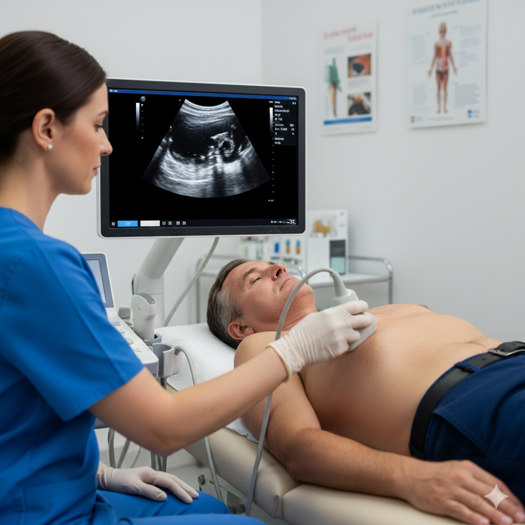
What is 2D/3D Echocardiography with Doppler?
2D/3D Echocardiography with Doppler is an advanced ultrasound-based heart scan that provides live images of the heart’s structure and movement.
- 2D Echocardiography creates flat cross-sectional images of the heart.
- 3D Echocardiography offers detailed three-dimensional visualization for improved accuracy.
- Doppler Imaging measures blood flow speed and direction through the heart and blood vessels.
Together, these technologies help cardiologists evaluate heart performance, detect abnormalities early, and plan appropriate treatment.
Applications of 2D/3D Echocardiography with Doppler
Heart Valve Assessment
This test accurately detects valve narrowing, leakage, and other valve disorders by evaluating blood flow and valve movement in real time.
Evaluation of Heart Function
2D/3D Echo with Doppler helps measure pumping efficiency, heart muscle strength, and chamber function, which is important in heart failure management.
Detection of Congenital Heart Disease
The test identifies structural defects present since birth, including holes in the heart and abnormal blood vessel connections.
Diagnosis of Cardiomyopathy
It helps diagnose thickened or weakened heart muscles and assesses the severity of cardiomyopathy conditions.
Guidance During Cardiac Procedures
Real-time imaging assists doctors during angioplasty, valve repair, pacemaker implantation, and other interventional cardiac procedures.
Blood Flow Analysis
Doppler technology evaluates blood circulation through heart chambers and arteries, helping detect blockages or abnormal flow patterns.
Advantages of 2D/3D Echocardiography with Doppler
Non-Invasive and Safe
The procedure is completely non-surgical and painless, making it safe for patients of all ages.
Real-Time Heart Imaging
It provides live visualization of heart movement, valve function, and blood circulation for accurate diagnosis.
No Radiation Exposure
Unlike CT scans or X-rays, echocardiography uses sound waves and does not expose patients to harmful radiation.
Highly Accurate Diagnosis
3D imaging and Doppler technology improve accuracy in detecting heart abnormalities and planning treatment.
Quick and Convenient Procedure
The test is usually completed within 20–40 minutes and does not require lengthy preparation or hospitalization.
Cost-Effective Cardiac Evaluation
Compared to advanced imaging methods like MRI, this test offers detailed cardiac assessment at a more affordable cost.