- 19, Opp. Ramkrishna Math, Rahate Colony Road, Dhantoli, Nagpur -12, +91 9413564868

Peripheral Angiography & Interventions (DVT)
Peripheral angiography and vascular interventions are minimally invasive procedures used to diagnose and treat problems affecting blood vessels outside the heart, especially those supplying the legs. These procedures can help identify narrowed or blocked arteries and guide appropriate treatment.
Deep Vein Thrombosis (DVT) is different from arterial blockage. It occurs when a blood clot develops in a deep vein, usually in the leg. Depending on the severity and location of the clot, treatment may involve medicines or, in selected cases, catheter-based intervention.
At Rhythm Heart & Critical Care Hospital, Nagpur, patients with suspected peripheral vascular problems can receive appropriate evaluation and interventional treatment based on their individual condition.
What Is Peripheral Angiography?
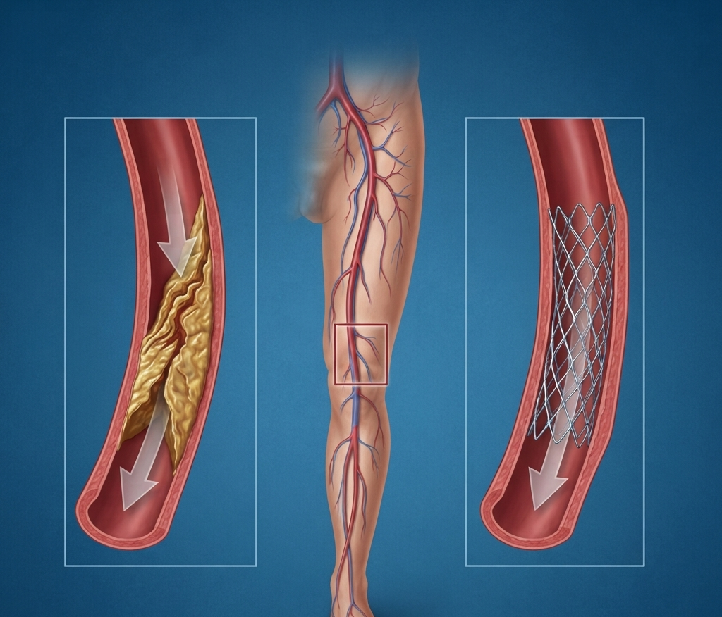
Peripheral angiography is an imaging test used to examine the blood vessels of the legs and other peripheral areas. A thin catheter is guided into the blood vessel and contrast dye is injected, allowing the doctor to identify narrowing, blockages, or abnormal blood flow on X-ray imaging.
If a significant blockage is found, the cardiologist may consider peripheral angioplasty, stenting, or another suitable intervention.
What Is DVT?
Deep Vein Thrombosis (DVT) occurs when a blood clot forms in a deep vein, most commonly in the leg. It may cause:
- Swelling in one leg
- Pain or tenderness
- Warmth around the affected area
- Red or discolored skin
DVT should not be ignored because, in some cases, the clot can travel to the lungs and cause a serious complication called pulmonary embolism.
When May Peripheral Intervention Be Needed?
A peripheral intervention may be considered when a blood vessel has significant narrowing or blockage that is affecting circulation.
Depending on the condition, treatment may include:
- Peripheral angioplasty
- Stent placement
- Catheter-based treatment in selected vascular conditions
- Medicines and monitoring
- Further vascular investigations
The appropriate treatment is decided after reviewing symptoms, imaging findings, and the patient’s overall health.
How Is Peripheral Angiography Performed?
1. Initial Evaluation
The doctor reviews the patient’s symptoms, medical history, medications, and relevant diagnostic reports.
2. Catheter Placement
A thin catheter is inserted through an appropriate blood vessel, depending on the area being examined and the planned procedure.
3. Contrast Imaging
Contrast dye is introduced through the catheter while X-ray imaging is performed to visualize the blood vessels.
4. Identifying the Problem
The images help the cardiologist identify narrowing, blockage, or reduced blood flow in the peripheral vessels.
5. Intervention When Required
If treatment is necessary, angioplasty, stenting, or another appropriate catheter-based procedure may be performed during the same session when clinically suitable.
Benefits of Peripheral Angiography & Interventions
- Helps identify peripheral blood vessel blockages
- Provides detailed information about blood circulation
- Supports accurate treatment planning
- May restore blood flow in selected patients
- Can avoid the need for more invasive treatment in appropriate cases
- Allows targeted treatment of certain vascular blockages
Risks and Precautions
Peripheral angiography and interventions are invasive procedures and may have potential risks such as bleeding or bruising at the catheter site, blood vessel injury, infection, or reaction to contrast dye.
Before the procedure, inform your doctor about kidney disease, diabetes, allergies, blood-thinning medicines, and any previous reaction to contrast material.
Precautions After Peripheral Intervention
1. Take Medicines as Prescribed
Take all prescribed medicines regularly. Do not stop blood-thinning or other cardiac medicines without discussing it with your doctor.
2. Take Care of the Catheter Site
Keep the access area clean and check for unusual bleeding, swelling, increasing pain, or discharge.
3. Avoid Heavy Physical Activity
Avoid strenuous exercise and heavy lifting for the period recommended by your doctor after the procedure.
4. Stay Hydrated if Permitted
If your doctor has not restricted your fluid intake, adequate hydration may help your body eliminate the contrast material used during angiography.
5. Watch for Changes in the Leg
Report new or worsening swelling, pain, numbness, unusual coldness, or changes in skin color of the affected limb.
6. Follow Up With Your Doctor
Attend scheduled follow-ups so your doctor can assess blood circulation, recovery, medicines, and the need for further treatment.
Why Choose Rhythm Heart & Critical Care Hospital?
Rhythm Heart & Critical Care Hospital provides comprehensive cardiac and interventional care in Nagpur, with facilities for advanced catheter-based procedures. The hospital’s published information also highlights experience with peripheral angioplasties as part of its interventional cardiology services.
The focus is on appropriate diagnosis, careful procedural planning, and individualized treatment rather than using the same approach for every patient.