- 19, Opp. Ramkrishna Math, Rahate Colony Road, Dhantoli, Nagpur -12, +91 9413564868
Intravascular Imaging (IVUS, OCT) in Nagpur
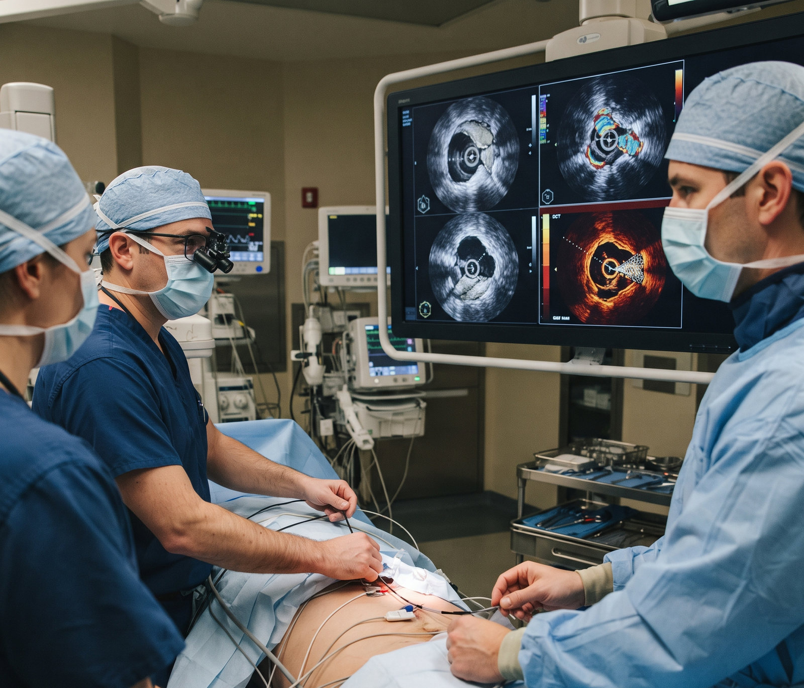
Intravascular Imaging is an advanced cardiac imaging technique used to examine the inside of coronary arteries in detail during selected cardiovascular procedures. Unlike conventional angiography, which provides an outline of the blood vessels, intravascular imaging uses specialized catheters to create detailed images of the artery from within. The two commonly used technologies are Intravascular Ultrasound (IVUS) and Optical Coherence Tomography (OCT).
These techniques can help cardiologists assess plaque, understand the size and condition of coronary arteries, guide stent placement, and check whether a stent has expanded and positioned properly. IVUS and OCT in Nagpur may be particularly useful during complex coronary interventions where additional information is required for accurate treatment planning.
Uses of Intravascular Imaging (IVUS, OCT)
A cardiologist may recommend IVUS or OCT during selected coronary procedures for various reasons, including:
- Assessing Coronary Artery Blockages: Helps provide detailed information about the size, location, and characteristics of a narrowed coronary artery.
- Evaluating Plaque: Can help identify plaque burden and certain features of plaque within the artery.
- Planning Stent Placement: Provides information about vessel dimensions that can assist in selecting an appropriately sized stent.
- Optimizing Stent Deployment: Helps evaluate stent expansion and apposition after placement.
- Assessing Complex Coronary Lesions: May provide additional information when coronary anatomy is difficult to evaluate through angiography alone.
How Is Intravascular Imaging (IVUS, OCT) Performed?
1. Preparation:
- The patient is prepared for the planned coronary catheter-based procedure.
- The medical team reviews the patient’s medical history, medications, allergies, and relevant test reports.
- Local anesthesia is generally used at the catheter insertion site.
- The cardiologist explains the procedure and determines whether IVUS or OCT is appropriate.
2. Introducing the Imaging Catheter:
- A thin catheter is carefully introduced into a blood vessel, commonly through the wrist or groin, depending on the procedure.
- The catheter is guided toward the coronary artery using imaging guidance.
- A specialized IVUS or OCT imaging catheter is then positioned in the area that needs detailed assessment.
3. Capturing Images:
- IVUS uses ultrasound waves to create cross-sectional images of the coronary artery.
- OCT uses near-infrared light to generate highly detailed images of structures within the vessel.
- The cardiologist reviews the images to assess the artery, plaque, lesion characteristics, and other relevant findings.
4. Supporting the Coronary Procedure:
- The information obtained can help the cardiologist determine appropriate treatment strategies.
- During angioplasty, imaging may assist with stent sizing and positioning.
- After stent placement, IVUS or OCT can be used to evaluate stent expansion and apposition when clinically appropriate.
5. Completing the Procedure:
- Once the required images have been obtained, the imaging catheter is removed.
- The cardiology team continues to monitor the patient according to the overall procedure being performed.
- The findings are documented and considered along with angiography and other clinical information.
What Happens After Intravascular Imaging?
- The cardiologist reviews the images obtained during the procedure.
- Findings from IVUS or OCT are considered along with angiography, symptoms, medical history, and other cardiac investigations.
- If the imaging identifies an issue with a coronary stent, the cardiologist may make appropriate adjustments during the same procedure when required.
- The imaging findings can also assist in planning further treatment or follow-up care.
- Your cardiologist will explain the results and discuss the recommended treatment plan.
IVUS vs OCT: What Is the Difference?
Intravascular Ultrasound (IVUS)
IVUS uses ultrasound waves to produce images from inside the coronary artery. It can provide deeper visualization of the vessel and is useful for assessing vessel dimensions, plaque burden, and stent deployment.
Optical Coherence Tomography (OCT)
OCT uses near-infrared light and provides very high-resolution images. It can offer detailed visualization of structures such as stent struts and features close to the vessel wall.
The choice between IVUS and OCT depends on the patient’s coronary anatomy, clinical condition, and the specific information required by the interventional cardiologist.
Benefits of IVUS and OCT
- Provides detailed images from inside coronary arteries.
- Supports assessment of vessel size and plaque characteristics.
- Helps with appropriate stent sizing and placement.
- Allows evaluation of stent expansion and apposition.
- Can provide additional information during complex coronary interventions.
- Supports more informed procedural decision-making.
- May help identify certain stent-related or vessel-related abnormalities.
Risks and Precautions
IVUS and OCT are minimally invasive techniques performed during catheter-based cardiovascular procedures. While they are generally considered useful and well-established imaging technologies, potential risks can be associated with the overall invasive procedure.
- There may be mild discomfort or bruising at the catheter insertion site.
- Rare complications can include bleeding, blood vessel injury, infection, or abnormal heart rhythm.
- Patients should inform the medical team about allergies, medications, and existing health conditions.
- The procedure should be performed by an experienced cardiovascular team with appropriate monitoring.
- Your cardiologist will explain the potential benefits and risks before the procedure.
Precautions After the Procedure
Follow Catheter-Site Care Instructions
Keep the catheter insertion area clean and follow the medical team’s instructions regarding activity and wound care.
Avoid Strenuous Activity Initially
Follow your cardiologist’s advice regarding lifting heavy objects, exercise, and other strenuous activities after the procedure.
Watch for Unusual Symptoms
Report significant swelling, persistent bleeding, severe pain, chest discomfort, breathing difficulty, or other concerning symptoms to your medical team promptly.
Take Prescribed Medicines
If medications such as antiplatelet or other cardiac medicines are prescribed, take them exactly as directed by your cardiologist.
Attend Follow-Up Appointments
Follow-up allows your cardiologist to review the procedure findings, assess your recovery, and determine whether additional treatment or monitoring is required.
Follow Your Cardiac Care Plan
Continue recommended lifestyle changes, medications, investigations, and follow-up care to support long-term cardiovascular health.
Intravascular Imaging (IVUS, OCT) in Nagpur
Intravascular Imaging with IVUS and OCT provides a closer look at the inside of coronary arteries during selected catheter-based cardiac procedures. IVUS uses ultrasound to assess vessel structure and dimensions, while OCT uses near-infrared light to provide highly detailed images of the vessel and stent.
These technologies can assist in evaluating coronary blockages, understanding plaque characteristics, planning stent placement, and checking stent expansion and apposition. They are particularly valuable when additional information is needed during complex coronary interventions.
If you have been advised coronary angiography, angioplasty, stent placement, or advanced coronary assessment, speak with an experienced cardiologist to understand whether IVUS or OCT in Nagpur may be appropriate for your treatment.
Book a cardiac consultation at Rhythm Heart And Critical Care Hospital for expert evaluation and personalized cardiovascular care.