- 19, Opp. Ramkrishna Math, Rahate Colony Road, Dhantoli, Nagpur -12, +91 9413564868
Coronary Angiography in Nagpur
Coronary Angiography in Nagpur
Coronary angiography is a specialized diagnostic procedure used to identify blockages, narrowing, or abnormalities in the coronary arteries that supply blood to the heart.
It is considered one of the most reliable methods for diagnosing coronary artery disease and determining the best treatment plan.
At Rhythm Heart & Critical Care Hospital, we provide advanced coronary angiography services using modern Cath Lab technology and experienced cardiologists to ensure accurate diagnosis and safe patient care.
Our hospital is equipped with state-of-the-art cardiac facilities to deliver comprehensive heart care under one roof.
What is Coronary Angiography?
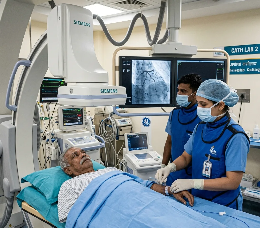
Coronary angiography is a minimally invasive diagnostic procedure used to examine the coronary arteries, which supply blood to the heart muscle. During the procedure, a thin flexible tube called a catheter is inserted through an artery in the wrist or groin and guided to the heart.
A special contrast dye is then injected into the coronary arteries, and X-ray images are taken to visualize blood flow. This allows cardiologists to detect blocked or narrowed arteries, reduced blood supply to the heart, and signs of coronary artery disease (CAD).
Coronary angiography is one of the most effective tests for identifying the cause of chest pain, assessing the risk of heart attack, and determining whether treatments such as angioplasty or bypass surgery may be required. Early diagnosis through coronary angiography helps ensure timely and appropriate heart care.
Why is Coronary Angiography Done?
A cardiologist may recommend coronary angiography if you experience:
- Chest pain or angina
- Shortness of breath
- Abnormal ECG or TMT results
- Suspected coronary artery disease
- Previous heart attack symptoms
- Unexplained heart problems
- Need for angioplasty or stent planning
Early diagnosis helps prevent severe heart complications and improves treatment outcomes.
Risks and Safety of Coronary Angiography
Coronary angiography is a safe and widely used procedure for diagnosing heart artery blockages. Although complications are rare, some patients may experience mild bleeding, bruising, temporary discomfort, or an allergic reaction to the contrast dye.
Serious issues such as infection or irregular heartbeat are uncommon. Most patients recover quickly and return home within 24 hours.
Coronary Angiography Procedure
The coronary angiography procedure generally involves the following steps:
1. Patient Preparation
The patient is asked to lie on the procedure table, and vital signs such as blood pressure, heart rate, and oxygen levels are monitored. Local anesthesia is administered to numb the insertion area.
2. Catheter Insertion
A thin, flexible catheter is carefully inserted through an artery in the wrist (radial artery) or groin (femoral artery) and guided toward the heart.
3. Catheter Positioning
Using real-time X-ray guidance, the cardiologist positions the catheter at the opening of the coronary arteries to ensure accurate imaging.
4. Contrast Dye Injection
A special contrast dye is injected through the catheter into the coronary arteries, making them visible on X-ray images.
5. X-Ray Imaging
Multiple X-ray images are captured from different angles to evaluate blood flow and identify any narrowing or blockages in the coronary arteries.
Results and Treatment Planning
The cardiologist reviews the angiogram images and discusses the findings with the patient. If significant blockages are detected, further treatment such as angioplasty or bypass surgery may be recommended.